Case presentation
A 27-year-old right-handed male presented with blurring of vision and squinting of his eyes upon waking up in the morning. He was well the evening before sleep. His symptoms were described as seeing blurred images and misaligned eyes with the right eye diverted inwardly. There was no speech difficulty, or facial or limb symptom. He had enjoyed good health without any known medical illness previously. His only significant history was being overweight with a smoking history of 10 pack-years.
He presented to our centre on day three of illness as his vision had worsened from blurred vision to diplopia. Upon assessment, there was right eye divergence on primary gaze. When instructed to look to the left side, there was impaired adduction of the right eye with nystagmus of the left eye. Other direction of gaze, accommodation and cranial nerves assessment were intact. All four limbs displayed normal tone, power and reflexes with bilateral flexor plantar responses. He was able to ambulate with ease.
His presentation was consistent with a right abducens nerve palsy and right internuclear ophthalmoplegia suggesting a lesion at the right dorsal pons. In view of his clinical presentation coupled with smoking risk factor, he was treated as posterior circulation stroke with aspirin and lipid-lowering agent. He was admitted for further work up and observation.
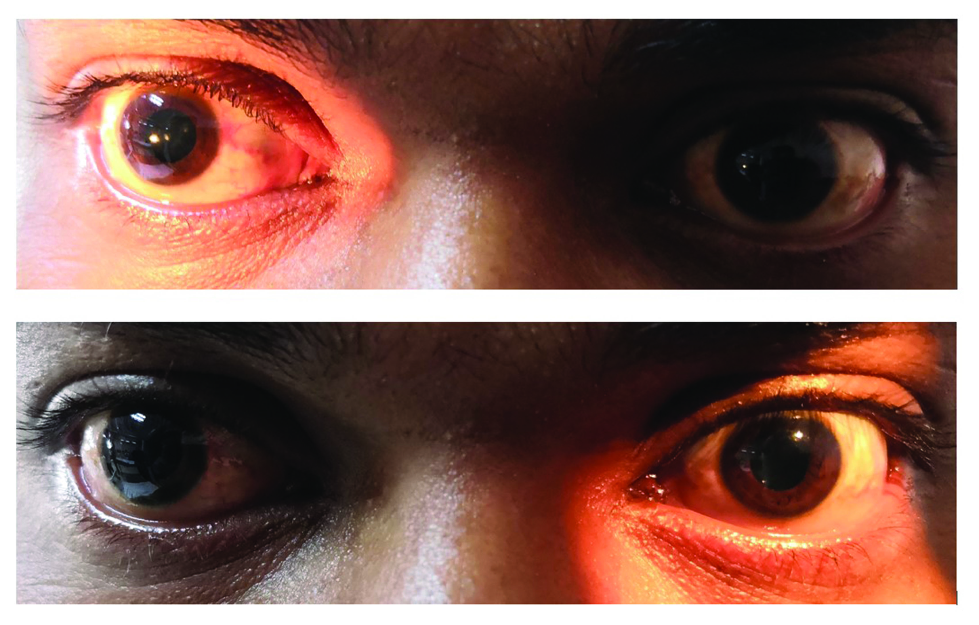
On day five of illness there were evolving signs. His extraocular movements were partially restricted at all directions of gaze accompanied by bilateral horizontal gaze evoked nystagmus. Nonfatigable unilateral partial ptosis was observed with both pupils dilated at 5 mm and appeared nonreactive (Figure 1). He swayed as he tried tandem walking. All his limb reflexes were present except for the left ankle, which was absent despite reinforcement.
Figure 1 Fixed dilated pupils at day five of illness

A plain CT of brain performed on admission did not show any hypodensities or intracranial bleed. An inpatient MRI with angiogram of the brain was reported as normal without any restricted diffusion to suggest acute stroke. No extracranial and intracranial arteries stenosis was seen in the angiogram. In view of the negative neuroimaging and evolving clinical signs, the initial diagnosis of stroke was unlikely. At one point, he was started on Pyridostigmine as empirical treatment of ocular myasthenia gravis without any clinical improvement seen. Nerve conduction study (including F waves), repetitive nerve stimulation test and single fibre electromyography at day seven of illness were normal.
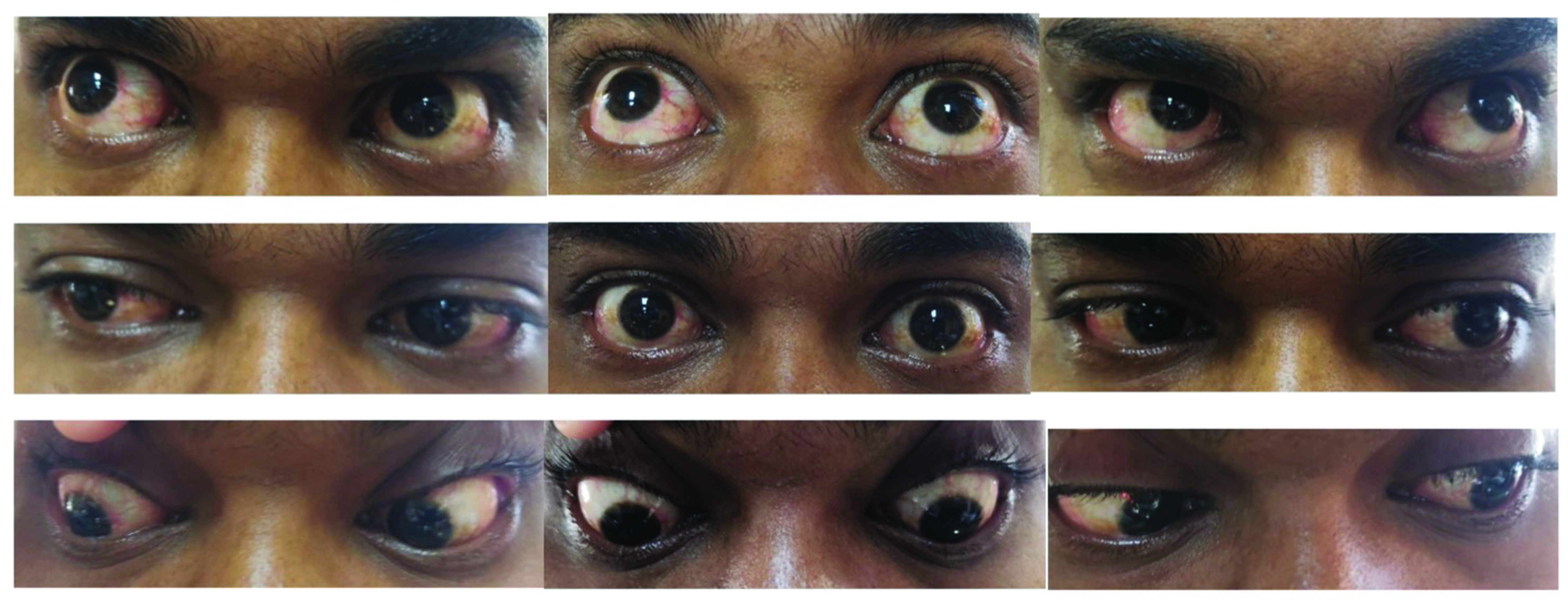
On day six of illness, he became more ataxic with worsening of ophthalmoplegia (Figure 2). All limb reflexes were absent despite reinforcement. A working diagnosis of Miller Fischer syndrome (MFS) was entertained at this point of time. He had one episode of vomiting with dyspepsia a week before the symptoms.
Figure 2 Nine-gaze view showing the ophthalmoplegia

He was treated with intravenous immunoglobulin 0.4 g/kg/day for 5 days as his symptom was presumed to be disabling (not able to ambulate independently). Unfortunately, diagnostic lumbar puncture was offered but rejected by patient. He showed gradual improvement and at day 10 of illness he was discharged with residual ataxia and ophthalmoplegia (modified Rankin scale of one). He recovered fully when seen in clinic 2 weeks after discharge. His pupils were back to baseline at 3 mm bilaterally and reactive. His 2-week post-treatment anti-GQ1b antibodies and anti-ACH receptor antibodies were negative.
Discussion
MFS is a variant of Guillain–Barré Syndrome (GBS) described by Charles Miller Fischer in 1956.1 The extramedullary portions of oculomotor, trochlear and abducens nerves contain rich amounts of GQ1b ganglioside that serve as the pathophysiological basis of MFS. Anti-GQ1b antibodies, which are present in 85–90% of all MFS cases, target this area causing the observed clinical features.2 The classical form of MFS consists of ophthalmoplegia, areflexia and ataxia. Forme fruste form has been described as exemplified by our case.3
The present case was a diagnostic challenge as during the initial presentation his symptomatology was still evolving. He was treated as posterior circulation stroke. As the signs still evolved, other differential diagnoses were considered. The partial ptosis seen at one point had made us think of ocular myasthenia gravis. The appearance of dilated, nonreactive pupils was an important sign in our case as pupillary involvement is not seen in ocular myasthenia gravis. Pupillary abnormalities are seen in MFS owing to antibody-mediated ciliary nerves and ganglion involvement.4 Anti-GQ1b antibodies were negative in our case probably because the serology was taken 2 weeks after treatment with intravenous immunoglobulin, ideally this investigation should be carried out prior to treatment. Hence the present case was unique given the evolving symptomatology, bilateral mydriasis and negative anti-GQ1b antibody status.
This case emphasises the need for ward admission for observation and work up as the initial presentation may be misleading. The likely diagnosis of MFS was only entertained after ruling out important differentials such as stroke and myasthenia gravis, accompanied by clinical improvement with intravenous immunoglobulin.
The role of disease-modifying treatment (immunoglobulin and plasma exchange) for MFS is less established compared with GBS.5 The indication for disease-modifying treatment includes nonambulatory cases, rapidly progressive cases as well as respiratory and bulbar involvement. We decided to give intravenous immunoglobulin (IVIG) for our case as his symptom was disabling.
The administration of IVIG hastened his recovery as he attained baseline status after 2 weeks of treatment.
Conclusion
The present case highlights that MFS, especially the forme fruste form, may cause diagnostic confusion with other neurological disorders, i.e. posterior circulation stroke and myasthenia gravis.
References
1 Fischer M. An unusual variant of acute idiopathic polyneuritis (syndrome of ophthalmoplegia, ataxia and areflexia). N Engl J Med 1956; 255: 57–65.
2 Chiba A, Kusunoki S, Obata H et al. Serum anti-GQ1b IgG antibody is associated with ophthalmoplegia in Miller Fisher syndrome and Guillain-Barré syndrome: clinical and immunohistochemical studies. Neurology 1993; 43: 1911.
3 Pritchard J. Review: what’s new in GBS? Postgrad Med J 2008; 84: 532–8.
4 Kaymakamzade B, Selcuk F, Koysuren A et al. Pupillary involvement in Miller Fisher syndrome. Neuroopthalmology 2013; 37: 111–5.
5 Verboon C, Doets AY, Galassi G et al. Current treatment practice of Guillain-Barré syndrome. Neurology 2019; 93: e1–e18.
