A personal Bell’s palsy and the ‘cheek puff’ sign
Some years ago, I suffered a Bell’s palsy, unpleasant in ways that were clinically educative. The experience was educative, too, in fostering an interest in Charles Bell and in the infamous surgeon-anatomist Robert Knox whose paths in life had, to my surprise, crossed.
It all began one morning with scrambled eggs. They seemed to have no taste and had an odd texture, like marble. They had been made no differently. Thinking no more of it, I drove to work. Straightening my tie in the mirror before student teaching, my face looked curiously impassive. I made to smile and only the right side of my mouth moved. I could not shut my left eye. It was a Bell’s palsy. This explained the scrambled eggs and the left earache I had had through the night. The teaching session that morning was unforgettable – for both parties!
Prednisolone 80 mg daily for three days, then seven days tapering off was prescribed. It was a weird experience. A Bell’s palsy may be regarded as a banal affliction but it is upsetting. Deprived of normal facial expression, it was disorientating to have to concentrate on the spoken word. Even speech could have been affected, orbicularis oris weakness interfering with the articulation of labial sounds, but I did not notice this. Involvement of three special senses: vision, hearing and taste, added to the weirdness. Embarrassing tears blurred and overflowed my sagging eyelid and the eye felt gritty. Loud noises were unpleasantly louder, the stapedius branch of the facial nerve not damping ossicle movements. Loss of sensation to much of the left half of my tongue from an inoperative chorda tympani branch explained the dysgeusia. High-dose corticosteroids further added to the strain, with insomnia and probably other subtle effects. Being on edge at my disfigurement did not help and my cheering habit of whistling – irritating to some! – was impossible. Not until something goes wrong does one appreciate how much normality is taken for granted.
I had the full house of clinical signs of a lower motor neurone facial nerve lesion except that, to my surprise, I could puff out my left cheek without trouble but not, separately, my right. Both could be puffed out together, but the left was fuller and the right easier to deflate. Everyone was surprised. Some months later, examining in the ‘Membership’, one of the cases was a man with a right-sided Bell’s palsy. I challenged my co-examiners on what they expected to find. All said weakness in blowing out the right cheek. Wrong: there was the same contralateral weakness I had found in myself.
Reflecting on this now, I think I have the answer. Books do not help. The names of authors ring down the years: Cecil & Loeb (now Goldman-Cecil), Harrison, Davidson, Hutchison & Hunter, Chamberlain, Garland & Phillips, Pappworth, MacLeod, the Oxford Textbook, Souhami & Moxham, Kumar & Clark, Jones & Tomson, Talley & O’Connor, McHardy et al. I also checked neurological texts, some venerable: Brain, Walton, Gowers, Kinnier Wilson, Walshe, Purves-Stewart, De Jong, Patten, Bickerstaff, Merritt, Bastron et al., Elliott et al. and Lindsay & Bone. I looked at a 1908 Osler and at various review articles. Less than half of all these publications mention puffing out the cheeks, some as an unqualified comment, some advising to look for asymmetry and/or to test resistance to deflation. Only three specify unambiguously which side cannot be puffed out.
How many readers would not expect it to be, as with all the other signs, the ipsilateral? The written contexts imply it, and Walshe, De Jong and a very recent US review article – the unambiguous three – affirm it: that it is the paralysed cheek that cannot be puffed out.1-3 But I had found the opposite and the explanation now seems obvious: that it was the inability of my paralysed left buccinator to contract and compress and flatten the cheek and thereby resist the passage of air into it that accounted for the seeming anomaly. I could separately blow out this unresisting left cheek because the right was ‘shut’ flat by the normal contraction of its unaffected buccinator, whereas puffing out the right cheek separately was impossible as air escaped into the unresisting left. Try blowing air alternately into each cheek and, though it is barely perceptible, each opposite buccinator can be felt alternately tensing. When I blew both out together, the fuller left cheek and easier to deflate right reflected the complete lack of tone in that left buccinator, though some air also escaped from the left side of my mouth. Only one text described, in part, a similar observation. Chamberlain’s Symptoms and Signs in Clinical Medicine states, without further elaboration, that ‘when the cheeks are puffed out the paralysed side balloons more than normal’.4 Paralysis of the buccinator may also allow food to lodge between the teeth and the cheek on the ipsilateral side and interfere with chewing.
I do not expect eponymous immortality for my cheek puff sign, what a colleague wittily suggested might be called the ‘paradoxical puff’. My observation is an inconsequential nicety. Bell’s palsy is easy to diagnose without it. Ironically, the idiopathic facial palsy which now bears his name was not amongst the cases he described to the Royal Society in his two seminal papers on the seventh cranial nerve in 1821 and 1829.5,6 Interestingly, he called this nerve the ‘respiratory nerve of the face’ and, recognising this might be confusing, elegantly explained why: that he regarded some of the facial muscles as accessory to exertion, such as their flaring of the nares. Interesting, too, he credited a branch of the trigeminal nerve as motor to the buccinator, mistakenly assuming it to be part of a coordinated process of mastication, a not unreasonable view. He did not mention puffing out the cheek. Although he was the first to demonstrate that facial palsy is due to a lesion of the seventh cranial nerve, the clinical condition had long been described by many of the medical greats of the first millennium, such as Celsus, Avicenna and Razi.7 Artefacts picturing facial palsy have been found in ancient Egypt, ancient Greece and Pre-Columbian America.8
Sir Charles Bell
But there is much more to Sir Charles Bell FRS (1774-1842) than his palsy. Born in Fountainbridge, Edinburgh, he went to the city’s high school and then to its university medical school, though did not take a degree, being trained in anatomy and surgery by his elder surgeon-anatomist brother, John.9 In 1799 he was admitted as a member of the Royal College of Surgeons of Edinburgh. He moved to London in 1804, was appointed surgeon to the Middlesex Hospital in 1810 and Professor of Physiology and Surgery at the new University of London in 1828. In 1836, he returned to Edinburgh to the Chair of Surgery.
He is credited with the idea that the brain is divided into different functional regions and that nerves are not single nerves serving various functions but are made up of bundles of different nerves with different functions and different origins in the brain, ‘united for convenience of distribution’.10 The latter observation came in part from his seminal animal experiments on the anterior and posterior spinal nerve roots. Some of his further conclusions were wrong or muddled, but his basic idea was a key contribution to neurophysiology. Some have likened its importance to that of Harvey’s discovery of the circulation of the blood, even arguing that Bell did not have Harvey’s advantage of pre-discovered facts upon which to reason, such as the presence of venous valves.9 The distinguished neurologist Charles-Édouard Brown-Séquard, writing 50 years after Bell’s idea, considered it largely responsible for subsequent progress in neurology.11
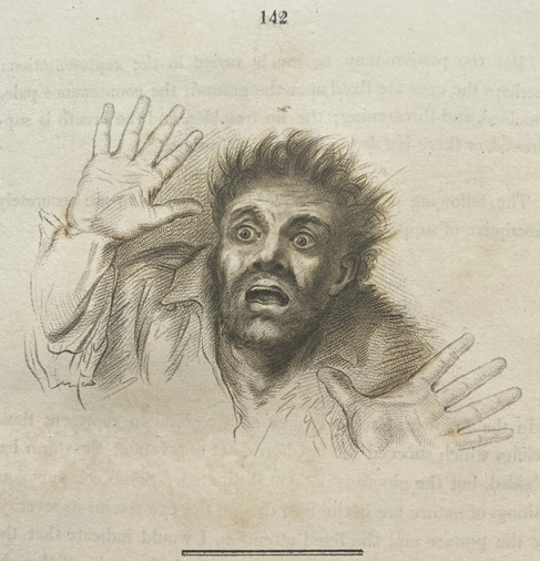
Bell was also a highly accomplished artist and published his self-illustrated Essays on the Anatomy of Expression in Painting in 1806 (Figure 1).
Figure 1 ‘Wonder, astonishment, fear, terror, horror, despair’. From Bell’s Essays on the Anatomy of Expression in Painting. London: Longman, Hurst, Rees & Orme; 1806. p.142

Darwin, in his The Expression of the Emotions in Man and Animals in 1872, credited Bell with having laid the scientific foundation for the study of expression and frequently quoted him, though his evolutionary ideas differed from Bell’s religious position. In 1833, Bell wrote the fourth Bridgewater Treatise, The hand; its mechanism and vital endowments, as evincing design, and illustrating the power, wisdom, and goodness of God, one of a series of texts written in support of natural theology. It is worth reading today whatever one’s beliefs. Full of comparative anatomy, it actually supports evolution.
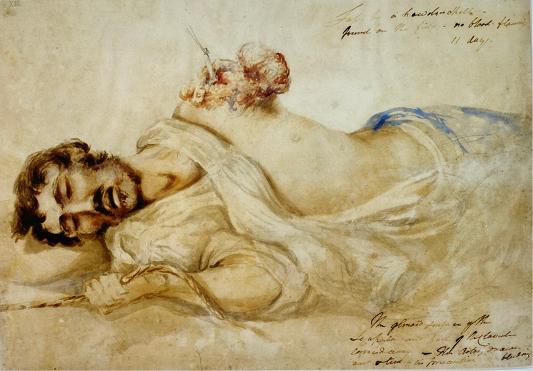
Bell used his artistic talents to paint the wounded when, as one of just a few civilian surgeons, he volunteered to help during the Napoleonic Wars. He treated wounded British troops returning from Corunna in 1809 and went to Belgium in 1815 where he worked tirelessly on allied and enemy casualties from Waterloo. In his letters, edited by his niece, he records operating incessantly for 13 hours in the day in one three-day period, his ‘arms powerless with the exertion of using the knife’, and finding that one and a half hours sleep in 24 was sufficient, when normally he needed eight.12 He adds that the French had ‘the most horrid wounds, left totally without assistance’. His paintings of the wounded at Corunna and Waterloo are among the best-ever artistic depictions of the mutilations of war (Figure 2).
Figure 2 ‘Left arm, with acromial end of the clavicle, and glenoid cavity of the scapula, carried off by cannon shot’ at Waterloo. From Crumplin MKH & Starling P. A Surgical Artist at War: The Paintings and Sketches of Sir Charles Bell 1809-1815. Edinburgh: Royal College of Surgeons; 2005. p. 80. (Reproduced by permission of the Trustees of the Army Medical Services Museum)

The Bell Collection
The Royal College of Surgeons of Edinburgh acquired 15 of these paintings when, in 1825, it bought Bell’s valuable, over 3,000 item collection of anatomical drawings and preparations, pathology specimens and other material for £3,000 (c £288,000 today). It was shipped to Leith on board the smack Robert Bruce then conveyed to Surgeons’ Square in spring wagons lent by the Artillery. The story is told in the College’s history of its museum.13 To my surprise, Robert Knox, surgeon-anatomist and brilliant teacher, who would be at the centre of the Burke and Hare, murders-for-cadavers scandal three years later, facilitated the sale, visiting Bell in London to assess the collection. He declined the College’s offer of £21 to defray expenses, declaring the visit his duty and of no inconvenience. It appears he made a second visit to supervise packing.9 He was soon after appointed Conservator of the College’s museum, where the collection today takes pride of place. Assiduously carrying out his duties, he was manoeuvred into resigning after the Burke and Hare affair.
Dr Robert Knox
Knox, as a 23-year-old recently qualified doctor, had also worked in Belgium in 1815 as an assistant surgeon and at some point, then or long after, claimed that only one of Bell’s amputation cases survived.14 This was out of an apparent total of 12, according to Bell’s biographers.9 If true, this would not necessarily have been due, wholly or partly, to poor surgery, for Bell was faced with having to perform secondary, delayed, amputation after sepsis had set in, a procedure with a much poorer prognosis than primary surgery in the field. Gangrene was also, according to Knox, rife in the Brussels hospital in which they both worked14 (and tetanus was common). Yet, while Bell is said to have had considerable skill as a civilian surgeon, a 92% mortality rate for secondary amputation was twice as high as was apparently then the case.9 It is not known, however, if there was any selection bias: whether Bell, by chance or design, was dealing with particularly ill cases. We do know that most of the injuries he dealt with were between two and three weeks old, very adequate time for serious infection, including septicaemia, to have set in. Nonetheless, whatever the explanation, 92% was a terrible mortality rate and Knox said that the people of Brussels spoke disparagingly of the ‘English medical department’.14
Is there any reason to question the number of amputations Bell carried out? His biographers do not provide evidence for their quote of 12 and it seems surprising he did not do more, considering how hard he worked. But he was a conservative surgeon and his biographers otherwise write with meticulous attention to detail and accuracy. Had he remarked on his arms being powerless from using the saw, one might be on stronger grounds for doubting the figure.
Can we believe Knox? He had a tendency to be arrogant towards professional colleagues and others. He sneered at the Bridgewater Treatises, undiplomatically referring to them as the ‘Bilgewater Treatises’.15 This would not have endeared him to Bell who was one of a number who opposed Knox’s application for an academic post at Edinburgh University in 1837. But by then Knox’s reputation was seriously tarnished by the Burke and Hare affair and his career in the city ruined, even though he had not been prosecuted as there was no evidence he knew the bodies he bought for dissection had been obtained by murder. He died in 1862, 20 years after Bell, and the provenance of the comment in his biography that ‘only one of Sir C. Bell’s [amputations] lived!’, which is the only identified source of the remark and written eight years after his death, is unclear. It may have been made long after Waterloo and perhaps influenced by Knox’s resentment at how he had been treated. However, in a fascinating book on fish and fishing, published in 1854, Knox writes that Charles Bell was the only person with whom he had never quarrelled, and adds ‘Peace be with him’.16 He also rather endearingly describes Bell once being seen by a riverside ‘stalking about strongly resembling the Ghost in “Hamlet”… cased to the eyes in waterproof!’.16 Taken in the round, it seems unlikely that he would have mischievously misrepresented Bell’s amputation results. Knox wrote six other books, including the controversial The Races of Men: a Fragment (1850) and A Manual of Artistic Anatomy, for the use of Sculptors, Painters and Amateurs (1852). He also published over a hundred scientific papers, many on anthropology and comparative anatomy.15
Bell himself planned a book on fishing, writing in some rough notes he had made: ‘By fishing, you contemplate nature; you are interested in the weather, in the winds that blow; in insects, their season, and their habits and propagation. And the fishes are a study… they have their time of rest, and of activity, and of feeding… your eye acquires a capacity for distant and minute objects; your hand, dexterity; your fingers, neatness. It is in fishing that you are brought to spots of secluded loveliness’.12
There was certainly much more to Charles Bell than his palsy and to Robert Knox than Burke and Hare. Knox was a more multidimensional and interesting character than popularly portrayed.
Conclusion
It is salutary that a doctor should be a patient at least once. It is a great leveller and cannot but help engender empathy in dealing with one’s own patients. If, as recounted here, the experience serves to stimulate curiosity to learn more – as in my case, of Bell and Knox – this is a bonus. They certainly deserve to be better known. Their biographies by Gordon-Taylor and Walls, and by Bates, to which references have been made, are excellent. 
Acknowledgements
I must pay tribute to Estela Dukan, Assistant Librarian at the Royal College of Physicians of Edinburgh, for her prompt, reliable and tireless help in responding to my requests for books and journal articles. Nothing is ever too much trouble for her. Thanks also to Dr Jacqueline Cahif, Archivist at the Royal College of Surgeons of Edinburgh, for her assistance. I am indebted to Dr Ian Ollerhead, dentist, for his advice and to Dr Brian Pentland, neurologist, for expanding my repertoire of neurology textbooks to consult.
References
1 Walshe FMR. Diseases of the Nervous System: Described for practitioners and students. Edinburgh: E & S Livingstone; 1940. p. 214. (Also in 9th edition, 1958. p. 292).
2 De Jong RN. The Neurological Examination. (4th ed). Hagerstown: Harper & Row; 1979. p. 186.
3 Reich SG. Bell’s palsy. Continuum (Minneap Minn) 2017; 23: 447-66.
4 Chamberlain EN. Symptoms and Signs in Clinical Medicine: An introduction to medical diagnosis. Bristol & London: Wright & Simpkin Marshall; 1936. p. 329. (Also in 12th edition, 1997. p. 229).
5 Bell C. On the nerves; giving an account of some experiments on their structure and functions, which lead to a new arrangement of the system. Phil Trans 1821; 111: 398-424.
6 Bell C. On the nerves of the face; being a second paper on that subject. Phil Trans 1829; 119: 317-30.
7 Pearce JMS. Early observations on facial palsy. J Hist Neurosci 2015; 24: 319-25.
8 Resende LA de L, Weber S. Peripheral facial palsy in the past: contributions from Avicenna, Nicolaus Friedreich and Charles Bell. Arq Neuropsiquiatr 2008; 66: 765-9.
9 Gordon-Taylor G, Walls EW. Sir Charles Bell: His Life and Times. Edinburgh: Livingstone; 1958. p. 8; p. 104; p. 140; p. 94.
10 Bell C. Idea of A New Anatomy of the Brain: submitted for the observations of his friends. London: Strahan & Preston; 1811. Reprinted in reference 9, pp. 218-31.
11 Brown-Séquard CE. Course of Lectures on the Physiology and Pathology of the Central Nervous System. Delivered at the Royal College of Surgeons of England in May, 1858. Philadelphia: Collins; 1860. p. 3.
12 [Bell M (ed)]. Letters of Sir Charles Bell, selected from his correspondence with his brother George Joseph Bell. London: Murray; 1870. pp. 246-7; pp. 429-30.
13 Tansey V, Mekie DEC. The Museum of the Royal College of Surgeons of Edinburgh. Edinburgh: Royal College of Surgeons; 1982. pp. 7-13; 16-18.
14 Lonsdale H. A Sketch of the Life and Writings of Robert Knox the Anatomist. London: Macmillan; 1870. p. 9.
15 Bates AW. The Anatomy of Robert Knox: Murder, Mad Science and Medical Regulation in Nineteenth-Century Edinburgh. Brighton: Sussex Academic Press; 2010. pp. 113-4; pp. 205-8.
16 Knox R. Fish and Fishing in the Lone Glens of Scotland: with a History of the Propagation, Growth, and Metamorphoses of the Salmon. London: Routledge; 1854. p. 12.